Auscultation is one of the first clinical skills students learn and one of the hardest to master. Textbooks can describe a murmur, but recognizing it by ear takes repeated, deliberate listening. This guide covers the basics: where to listen, which side of the stethoscope to use, and how the most common heart and lung sounds differ. Numbers in brackets refer to the references at the end.
Before you listen
- Bare skin. Auscultation should not be done through clothing [5].
- Diaphragm for high-pitched sounds, bell for low-pitched sounds [3][4]. Low-pitched sounds such as S3 and the murmur of mitral stenosis are heard with the bell [2][3].
- Know the positions. Patients can be examined lying supine, in the left lateral decubitus position, and sitting up leaning forward [4]. Some sounds are only obvious in one of these positions (see below).
The cardiac listening areas
| Area | Location |
|---|---|
| Aortic | 2nd intercostal space, right sternal border [1] |
| Pulmonic | 2nd intercostal space, left sternal border [1] |
| Erb’s point | 3rd intercostal space, left sternal border [3] |
| Tricuspid | 4th intercostal space, left sternal border [1] |
| Mitral (apex) | 5th intercostal space, left midclavicular line [1] |

Aortic, pulmonic, tricuspid and mitral auscultation points (Erb’s point is not shown). Image: Theodore Dimitriou, CC BY-SA 3.0, via Wikimedia Commons.
Murmurs may also radiate, so they can be heard away from these areas [1]: the murmur of aortic stenosis may radiate to the carotid arteries, and mitral regurgitation to the left axilla [1][4].
Normal heart sounds: S1 and S2
S1 results from closure of the mitral and tricuspid valves; its mitral component is loudest at the apex [2]. S2 results from closure of the aortic and pulmonic valves; its aortic component is loudest at the right upper sternal border [2].
Tip: to tell them apart, feel the carotid pulse while you listen. S1 just precedes the carotid pulse and S2 follows it [4]. Because systole is shorter than diastole, S1 and S2 sound grouped together, with S1 first [4].
Splitting of S2
- Physiological split: on inspiration, increased venous return delays the pulmonic component (P2), so S2 splits; the split disappears on expiration [2].
- Persistent (wide) split: both components are heard throughout respiration and the split widens on inspiration. Causes include right bundle branch block, pulmonary hypertension, pulmonic stenosis, severe mitral regurgitation and VSD [2].
- Fixed split: the split is the same throughout respiration and is associated with atrial septal defect [1][2].
- Paradoxical split: the split is heard on expiration and disappears on inspiration. It occurs with aortic stenosis, hypertrophic obstructive cardiomyopathy or left bundle branch block [2].
Extra heart sounds: S3 and S4
- S3 occurs in early diastole, during passive filling of the left ventricle. It may be normal in children, pregnant women and well-trained athletes, but it is often a sign of systolic heart failure. It is low-pitched and is best heard with the bell at the apex, with the patient in the left lateral decubitus position [2].
- S4 occurs in late diastole, when atrial contraction pushes blood into a stiff (noncompliant) left ventricle. It is almost always abnormal and may indicate diastolic heart failure, active ischemia or left ventricular hypertrophy. Because it depends on atrial contraction, an S4 cannot occur in atrial fibrillation [2].
Common murmurs at a glance
| Murmur | Timing and character | Where / radiation |
|---|---|---|
| Aortic stenosis | Midsystolic, crescendo-decrescendo (“diamond-shaped”), high-pitched [1][3] | Right upper sternal border; may radiate to the carotids [1] |
| Mitral regurgitation | Holosystolic, high-pitched, blowing [3] | Apex; may radiate to the left axilla [1][3] |
| Aortic regurgitation | Early diastolic, decrescendo, soft and high-pitched [3] | Left sternal border, usually loudest at Erb’s point [1][3] |
| Mitral stenosis | Low-pitched diastolic murmur after an opening snap [3] | Apex [1][3] |

Phonocardiograms showing where each murmur falls in the cardiac cycle. Image: Madhero88, CC BY-SA 3.0, via Wikimedia Commons.
Positioning matters. The murmur of mitral stenosis is accentuated in the left lateral decubitus position [4]. Aortic regurgitation is best heard with the patient sitting up, leaning forward, at end-expiration [3].
Normal breath sounds
- Vesicular (the normal lung sound): soft, low-pitched and rustling. Inspiration lasts longer than expiration, about 2:1, with no pause between them [5].
- Bronchovesicular: intermediate in intensity and pitch, with inspiration and expiration of similar length. Normally heard anteriorly over the 1st and 2nd intercostal spaces and posteriorly between the scapulae; it is abnormal in other locations [5].
- Bronchial: loud, hollow and high-pitched, with expiration longer than inspiration and a distinct pause between them. Normally heard over the manubrium; bronchial breath sounds with higher-frequency components occur in consolidation [5].
Technique: ask the patient to take deep breaths through the open mouth. Using the diaphragm, start anteriorly at the apices and move downward, then do the same on the back. Listen to at least one full respiratory cycle at each site, and always compare symmetrical points on each side [5].
Adventitious lung sounds
| Sound | What it sounds like | Notes |
|---|---|---|
| Fine crackles | Discontinuous; soft, higher-pitched, many per breath [5] | Seen in interstitial lung disease and early congestive heart failure [5] |
| Coarse crackles | Discontinuous; loud, low-pitched, fewer per breath [5] | Seen in chronic bronchitis and severe pulmonary edema. Early inspiratory crackles in COPD are usually due to secretions in large airways and disappear on coughing [5] |
| Wheeze | Continuous, musical, high-pitched; most often expiratory [5] | Not synonymous with asthma; found in a variety of conditions [5] |
| Rhonchi | Continuous, musical, low-pitched, with a snoring quality [5] | Best heard over the chest wall [5] |
| Stridor | Loud, high-pitched, musical; mainly inspiratory [5] | Produced by upper airway obstruction; louder over the neck than the chest wall [5] |
| Pleural rub | Non-musical, grating, creaky or leathery; in both phases of breathing [5] | Caused by inflamed pleural surfaces rubbing together; important to distinguish from crackles [5] |
How to train your ear
- Learn the normal first. Get comfortable with S1, S2 and vesicular breath sounds before moving on to abnormal ones.
- Listen for one thing at a time. At each area, focus on S1, then S2, then systole, then diastole, instead of trying to hear everything at once.
- Compare sounds side by side. Many sounds are easy to confuse, such as S3 and S4, fine and coarse crackles, or wheeze and stridor. Listening to them back to back makes the difference clearer.
- Test yourself. Naming a sound without seeing the label shows you what you actually recognize.
Practice with Lung Heart Sounds
Lung Heart Sounds is an auscultation trainer with 23 heart sounds and 9 lung sounds, built for medical, nursing and paramedic students, residents, and anyone preparing for OSCEs, clinical rotations, or cardiology and respiratory exams.
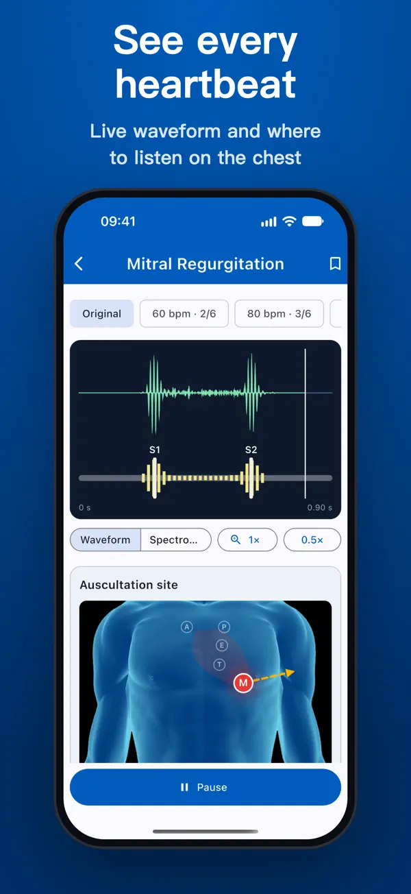
- See every heartbeat: a live phonocardiogram with S1 and S2 marked, a spectrogram view, and 0.5× playback to pick out splits, clicks and soft murmurs.
- Know where to listen: a chest diagram highlights the aortic, pulmonic, Erb’s point, tricuspid and mitral areas, with arrows showing where each murmur radiates and tips on bell or diaphragm, patient position and breathing.
- Heart sounds: normal heart sound, split S1, physiological, wide and paradoxical split S2, S3, S4, summation and quadruple gallop, the major valve murmurs, ASD, VSD, PDA, pericardial friction rub and pericardial knock.
- Lung sounds: vesicular, bronchovesicular and bronchial breath sounds, fine and coarse crackles, wheeze, rhonchi, stridor and pleural rub.
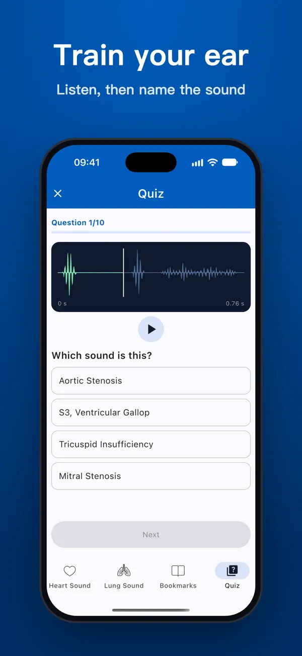
- Quiz mode: 10 questions per round mixing heart and lung sounds, with bookmarks to build your own review list.
- Works offline, with no account, no sign-up and no ads. Headphones are recommended, because low-pitched sounds such as S3, S4 and mitral stenosis are hard to hear on phone speakers. Available in English, Tiếng Việt, Español, 中文 and Portuguese.
Download on iOS: Lung Heart Sounds on the App Store
Download on Android: Lung Heart Sounds on Google Play
Disclaimer: this article is for educational purposes only and is not a substitute for formal clinical training or professional medical advice. Lung Heart Sounds is not a medical device and must not be used to diagnose or treat any condition.
References
- Thomas SL, Heaton J, Makaryus AN. Physiology, Cardiovascular Murmurs. In: StatPearls. Treasure Island (FL): StatPearls Publishing; last update July 17, 2023. https://www.ncbi.nlm.nih.gov/books/NBK525958/
- Lome S. Heart Sounds Topic Review. Healio, Learn the Heart. https://www.healio.com/cardiology/learn-the-heart/cardiology-review/topic-reviews/heart-sounds
- Lome S. Heart Murmurs Topic Review. Healio, Learn the Heart. https://www.healio.com/cardiology/learn-the-heart/cardiology-review/topic-reviews/heart-murmurs
- University of Washington School of Medicine. Techniques: Heart Sounds & Murmurs Exam. Physical Diagnosis Skills. https://depts.washington.edu/physdx/heart/tech.html
- Sarkar M, Madabhavi I, Niranjan N, Dogra M. Auscultation of the respiratory system. Ann Thorac Med. 2015;10(3):158–168. doi:10.4103/1817-1737.160831. https://pmc.ncbi.nlm.nih.gov/articles/PMC4518345/