Crackles and wheezes get most of the attention, but often the first thing you notice with the stethoscope on the chest is simpler: are the breath sounds there, and are they as loud as they should be? Diminished or absent breath sounds can point to obstructed airways, fluid around the lung or a misplaced breathing tube. Research shows when this simple finding helps, and when it misleads. Numbers in brackets refer to the references at the end.

Photo: Pavel Danilyuk, via Pexels.
Breath sound intensity and airflow obstruction
Pardee and colleagues asked four examiners to perform standardized physical examinations on patients who had just undergone tests of ventilatory function. The examiners graded the loudness of breath sounds during deep inspiration from 0 to 4 in six areas of the chest, giving a total score from 0 to 24 [1].
- The breath sound score correlated significantly with the percentage of predicted FEV1 for all four examiners, and the differences between examiners were not statistically significant.
- Grading breath sound loudness was a poor screening test for mild ventilatory abnormality.
- But normal breath sounds nearly excluded a severe reduction in FEV1, and definitely reduced breath sounds were strong evidence of obstructive pulmonary disease.
What this means: loudness is a rough gauge. It will not catch mild obstruction, but at the extremes, clearly normal or clearly reduced, it carries real information.
Diminished breath sounds in COPD
Badgett and colleagues studied 92 outpatients who smoked or had a history of COPD. Four internists, blinded to all other results, examined each patient for 12 physical signs, and the patients also had peak flow and spirometry. Fifteen patients (16%) had moderate COPD [2].
- Two items from the history, a previous diagnosis of COPD and smoking of 70 or more pack-years, diagnosed COPD with a sensitivity of 40% and a specificity of 100%.
- Diminished breath sounds was the only physical sign that added to the history, raising the mean sensitivity to 67% with a mean specificity of 98%.
- Patients with none of these three findings had a 3% prevalence of COPD.
The authors concluded that diminished breath sounds were the best predictor of moderate COPD, and that COPD was very unlikely with a normal history and physical examination [2].
A study with chest wall microphones adds a nuance. Ishimatsu and colleagues recorded breath sounds in 20 patients with stable COPD and 20 healthy controls. During resting breathing, breath sounds were actually louder in the COPD group. Only during deep breathing was the inspiratory sound diminished over the upper and middle lung fields, which the authors linked to reduced airflow [3].
What this means: “diminished breath sounds” is a finding of deep breaths. Both Pardee and Badgett assessed it in a standardized examination, and the acoustic study suggests that quiet tidal breathing may not show it at all.
Pleural effusion: the stethoscope is not the best tool
A JAMA Rational Clinical Examination review by Wong and colleagues looked for prospective studies that compared physical examination with radiographic confirmation of pleural effusion. Only 5 studies met the inclusion criteria (934 patients) [4].
- Eight examination manoeuvres were evaluated, including breath sounds, crackles, pleural friction rub, vocal resonance, chest expansion, tactile vocal fremitus and percussion.
- Dullness to conventional percussion was the most accurate finding for diagnosing pleural effusion (positive likelihood ratio 8.7).
- The absence of reduced tactile vocal fremitus made an effusion less likely (negative likelihood ratio 0.21).
The authors concluded that, based on the limited number of studies, dullness to percussion and tactile fremitus are the most useful findings, and that a dull chest still needs a chest radiograph to confirm the diagnosis [4].
What this means: when you hear quiet breath sounds at a lung base, do not stop at the stethoscope. Percuss and feel for fremitus as well.
Is the tube in the right place? Auscultation after intubation
Listening to both sides of the chest is a classic check after a breathing tube is placed. Sitzwohl and colleagues tested it in a randomised blinded trial of 160 adults having elective surgery. With a fibreoptic bronchoscope, the tube was placed either in the trachea or deliberately in the right main bronchus, and first-year residents and experienced anaesthetists then judged its position by one of four bedside methods [5].
- First-year residents missed endobronchial intubation by auscultation in 55% of cases and did significantly worse with this test than experienced anaesthetists.
- Auscultation had a sensitivity of 65% and observation of chest movements 43%, compared with 88% for tube insertion depth and 100% for all three methods combined.
- The four methods had the same specificity for confirming correct tracheal placement.
The authors concluded that less experienced clinicians should rely more on tube insertion depth than on auscultation, and that the best results came from combining tube depth, auscultation of the lungs and observation of symmetrical chest movements [5].
What this means: equal breath sounds on both sides are reassuring, but on their own they miss a tube in the right main bronchus too often, especially in inexperienced hands.
Critically ill patients: where auscultation falls short
Lichtenstein and colleagues compared auscultation, bedside chest radiography and lung ultrasound with thoracic CT in 32 ventilated patients with acute respiratory distress syndrome (ARDS) and 10 healthy volunteers, across 384 lung regions [6]:
| Diagnostic accuracy | Auscultation | Chest radiography | Lung ultrasound |
|---|---|---|---|
| Pleural effusion | 61% | 47% | 93% |
| Alveolar consolidation | 36% | 75% | 97% |
| Alveolar-interstitial syndrome | 55% | 72% | 95% |
The authors concluded that lung ultrasound is highly sensitive, specific and reproducible at the bedside in ARDS [6]. In ventilated patients, the stethoscope is a starting point, not the answer.
Lessons for listening to breath sounds
- Judge loudness on deep breaths. Reduced intensity showed up during deep inspiration, not quiet breathing [1][3].
- Trust the extremes. Clearly normal breath sounds make severe obstruction unlikely; clearly reduced ones support it. Mild changes tell you little [1].
- Combine the finding with the history. In smokers, diminished breath sounds was the only physical sign that added to the history of COPD and smoking [2].
- Add percussion and fremitus for a suspected effusion. They were the most useful examination findings in the review [4].
- Never rely on auscultation alone to check a breathing tube. Combine it with tube depth and chest movement [5].
- Know when to reach for imaging. In critically ill patients, auscultation was the least accurate tool for consolidation [6].
Practice with Lung Heart Sounds
Lung Heart Sounds is an auscultation trainer with 23 heart sounds and 9 lung sounds. Recognizing an abnormal breath sound starts with knowing what normal vesicular, bronchovesicular and bronchial breath sounds sound like.
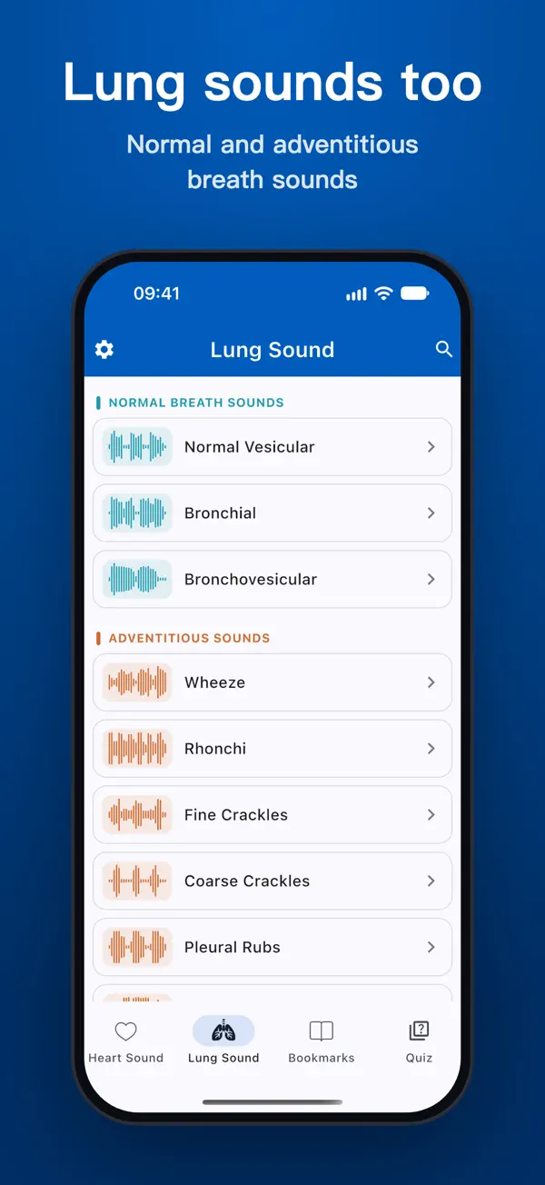
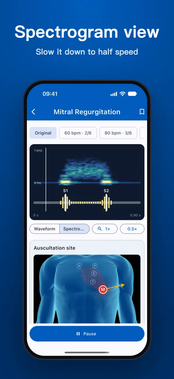
- 9 lung sounds: normal vesicular, bronchovesicular and bronchial breath sounds, plus fine crackles, coarse crackles, wheeze, rhonchi, stridor and pleural rub.
- See what you hear: a live waveform and a spectrogram view, and half-speed (0.5×) playback for any sound.
- Practical tips for each sound: diaphragm or bell, patient position and how the sound changes with breathing.
- Test yourself: quiz rounds of 10 questions mix heart and lung sounds and use different recordings each time. Bookmark sounds to build your own review list.
- Works offline, with no account and no ads. Headphones are recommended.
Download on iOS: Lung Heart Sounds on the App Store
Download on Android: Lung Heart Sounds on Google Play
Read more: our practical guide to heart and lung auscultation (including normal breath sounds and technique) and what research says about crackles, wheezes and rhonchi.
Disclaimer: this article is for educational purposes only and is not a substitute for formal clinical training or professional medical advice. The studies cited did not evaluate Lung Heart Sounds. The app is not a medical device and must not be used to diagnose or treat any condition.
References
- Pardee NE, Martin CJ, Morgan EH. A test of the practical value of estimating breath sound intensity. Breath sounds related to measured ventilatory function. Chest. 1976;70(3):341–344. doi:10.1378/chest.70.3.341. PMID 954460
- Badgett RG, Tanaka DJ, Hunt DK, Jelley MJ, Feinberg LE, Steiner JF, et al. Can moderate chronic obstructive pulmonary disease be diagnosed by historical and physical findings alone? Am J Med. 1993;94(2):188–196. doi:10.1016/0002-9343(93)90182-o. PMID 8430714
- Ishimatsu A, Nakano H, Nogami H, Yoshida M, Iwanaga T, Hoshino T. Breath sound intensity during tidal breathing in COPD patients. Intern Med. 2015;54(10):1183–1191. doi:10.2169/internalmedicine.54.3161. PMID 25986254
- Wong CL, Holroyd-Leduc J, Straus SE. Does this patient have a pleural effusion? JAMA. 2009;301(3):309–317. doi:10.1001/jama.2008.937. PMID 19155458
- Sitzwohl C, Langheinrich A, Schober A, Krafft P, Sessler DI, Herkner H, et al. Endobronchial intubation detected by insertion depth of endotracheal tube, bilateral auscultation, or observation of chest movements: randomised trial. BMJ. 2010;341:c5943. doi:10.1136/bmj.c5943. https://pmc.ncbi.nlm.nih.gov/articles/PMC2977961/
- Lichtenstein D, Goldstein I, Mourgeon E, Cluzel P, Grenier P, Rouby JJ. Comparative diagnostic performances of auscultation, chest radiography, and lung ultrasonography in acute respiratory distress syndrome. Anesthesiology. 2004;100(1):9–15. doi:10.1097/00000542-200401000-00006. PMID 14695718