A heart murmur or an extra heart sound is often the first clue to valve disease or heart failure, and learning to hear them is a classic part of medical training. But how much can the stethoscope really tell you? Research on cardiac auscultation gives a mixed but useful answer: some findings carry real diagnostic weight, skill clearly depends on experience, and the stethoscope works best when you know its limits. Numbers in brackets refer to the references at the end.

Photo: Antoni Shkraba, via Pexels.
Systolic murmurs: which features matter?
A review in JAMA’s Rational Clinical Examination series pooled the evidence on the clinical examination for abnormal systolic murmurs [1]. Most of the studies used cardiologists as examiners. Key findings:
- Agreement on whether a systolic murmur is present was only fair in the clinical setting (κ 0.30–0.48).
- For ruling in aortic stenosis, the most useful findings were a slow rate of rise of the carotid pulse (positive likelihood ratio 2.8–130), a murmur with mid to late peak intensity (8.0–101) and a decreased intensity of the second heart sound (3.1–50).
- For ruling out aortic stenosis, the most useful finding was the absence of murmur radiation to the right carotid artery (negative likelihood ratio 0.05–0.10).
- Smaller, lower-quality studies suggested that cardiologists can accurately rule in and rule out mitral regurgitation, tricuspid regurgitation, hypertrophic cardiomyopathy and echocardiographic mitral valve prolapse.
The authors concluded that the examination by cardiologists is accurate for detecting various causes of abnormal systolic murmurs, and called for studies of examiners who are not cardiologists [1].
What this means: it is not enough to hear “a murmur”. Its timing within systole, where it radiates and what happens to S2 are what turn a sound into a diagnosis.
Screening for valve disease in primary care: a sobering result
Gardezi and colleagues tested auscultation where it is often used: in primary care, on people without symptoms. Two experienced general practitioners examined 251 participants aged over 65 with no known valve disease, blinded to the echocardiography that all participants underwent [2].
- Previously unknown valve disease was common: mild disease in 170 of 251 participants (68%) and significant disease (at least moderate regurgitation or mild stenosis of any valve) in 36 of 251 (14%).
- The sensitivity of auscultation was 32% for mild and 44% for significant valve disease, with specificities of 67% and 69%.
- Likelihood ratios were not statistically significant in the overall cohort, but auscultation showed possible value in participants who were not overweight (body mass index below 25 kg/m2).
The authors concluded that auscultation is a poor screening tool for valve disease in asymptomatic primary care patients, and that easy access to echocardiography for patients with suggestive symptoms is likely a better strategy [2].
Innocent or pathological? Murmurs in children
Murmurs are common in children, and most are innocent. McCrindle and colleagues recorded the clinical impressions of five academic paediatric cardiologists for 222 consecutive children referred for a first evaluation of a heart murmur, then compared them with ECG and echocardiography [3]. The prevalence of heart disease was 33%.
- Clinical assessment identified pathological murmurs with a sensitivity of 92% and a specificity of 94%. When diagnostic uncertainty was treated as a reason for echocardiography, these rose to 97% and 98%. The lesions that were missed were only trivial or minor.
- Features independently associated with heart disease were a pansystolic murmur (odds ratio 54.0), an early or midsystolic click (8.35), a murmur of grade 3 or louder (4.84), a murmur heard best at the left upper sternal border (4.24), an abnormal second heart sound (4.09) and a harsh quality (2.37).
The authors suggested that these specific features may be of practical use to clinicians who are not cardiologists [3].
Gallops: the S3 and S4
The third (S3) and fourth (S4) heart sounds are low-pitched and soft, which makes them among the hardest sounds to hear. Research shows that they are worth the effort, with a caveat:
- The S3 is specific but not sensitive. In 90 patients undergoing heart catheterization, Marcus and colleagues detected the S3 and S4 with computerized phonocardiography. For detecting an elevated left ventricular end-diastolic pressure, a reduced ejection fraction or an elevated BNP, the S3 had sensitivities of 41%, 52% and 32% but specificities of 92%, 87% and 92%. The S4 had sensitivities of 46%, 43% and 40% and only moderate specificities of 80%, 72% and 78% [4].
- Hearing a useful S3 takes experience. In a second study of 100 patients, the same group compared the auscultation of physicians at four levels of training with phonocardiography. Findings of residents and interns showed no significant agreement with phonocardiography, while those of cardiology fellows (κ=0.37) and cardiology attendings (κ=0.29) did. The S3 had low sensitivity (13–52%) but high specificity (85–95%) for abnormal markers of left ventricular function, and its association with these markers improved with each level of experience [5].
What this means: not hearing an S3 does not rule out left ventricular dysfunction, but hearing a true S3 is a strong clue. How much the S3 adds in breathless patients with suspected heart failure is covered in our article on lung sounds.
Can artificial intelligence help the stethoscope?
Digital stethoscopes can record heart sounds and analyse them with algorithms:
- Chorba and colleagues trained a deep learning algorithm on more than 34 hours of heart sound recordings and tested it in 962 patients. It detected murmurs with a sensitivity of 76.3% and a specificity of 91.4%; when grade 1 murmurs were left out, sensitivity rose to 90.0%. At the appropriate auscultation site it detected moderate-to-severe or greater aortic stenosis (sensitivity 93.2%, specificity 86.0%) and mitral regurgitation (66.2% and 94.6%). The authors judged its performance comparable to expert cardiologists on the annotated subset of their data [6].
- In the TRICORDER trial, 205 UK primary care practices were randomised to use AI-enabled stethoscopes in routine care or to usual care. After 12 months, the detection of heart failure did not differ between the groups (incidence rate ratio 0.94). The use of the AI stethoscope was, however, independently associated with higher detection rates of heart failure, atrial fibrillation and valve disease, and the authors highlight the barriers to putting such tools into everyday practice [7].
Technology may extend the stethoscope, but it does not yet replace the clinician who knows where to listen and what the sounds mean.
Lessons for learning heart sounds
- Listen for the details, not just “a murmur”. Timing in the cardiac cycle, peak intensity, radiation and the second heart sound carry most of the diagnostic information [1][3].
- Know where each murmur radiates. For example, a murmur that does not radiate to the right carotid artery makes aortic stenosis unlikely [1].
- Learn the red flags in children: pansystolic, loud (grade 3 or more), harsh, a click or an abnormal S2 [3].
- Use the S3 to rule in, not to rule out. It is specific but insensitive at every level of experience [4][5].
- Know the limits. Auscultation is a poor screening test for valve disease in people without symptoms [2]; when in doubt, echocardiography decides.
Practice with Lung Heart Sounds
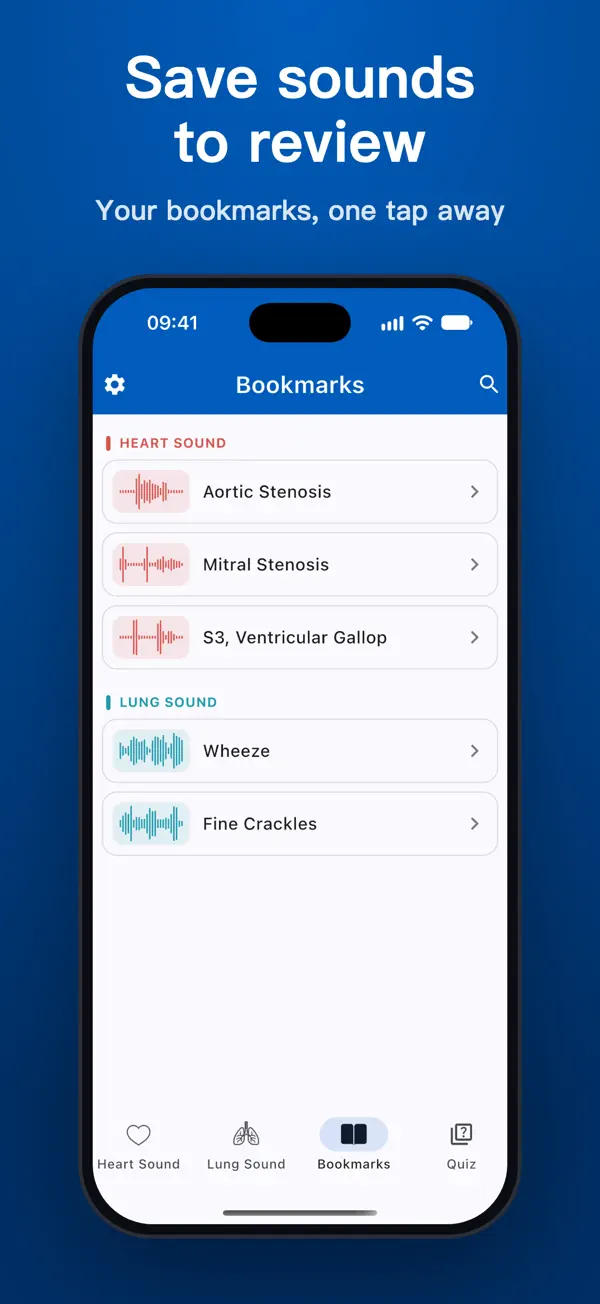
Lung Heart Sounds is an auscultation trainer with 23 heart sounds and 9 lung sounds, including the murmurs and gallops discussed in this article.
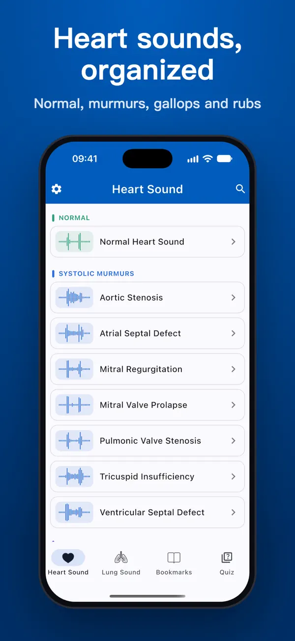
- 23 heart sounds: normal heart sound and splits; gallops (S3, S4, summation and quadruple gallop); murmurs including aortic stenosis, aortic insufficiency, mitral stenosis, mitral regurgitation and mitral valve prolapse; congenital and pericardial sounds.
- See what you hear: a live phonocardiogram with S1 and S2 marked, a spectrogram view to see the pitch and timing of murmurs, and half-speed (0.5×) playback to pick out splits, clicks and soft murmurs.
- Know where to listen: a chest diagram highlights the auscultation areas, loudest area first, with arrows showing where each murmur radiates, such as to the carotids or the axilla.
- Test yourself: quiz rounds of 10 questions use different recordings each time. Bookmark the sounds you confuse to build your own review list.
- Works offline, with no account and no ads. Headphones are recommended, since low-pitched sounds such as S3, S4 and mitral stenosis are hard to hear on phone speakers.
Download on iOS: Lung Heart Sounds on the App Store
Download on Android: Lung Heart Sounds on Google Play
Read more: our practical guide to heart and lung auscultation, what research says about getting better at auscultation and what research says about crackles, wheezes and rhonchi.
Disclaimer: this article is for educational purposes only and is not a substitute for formal clinical training or professional medical advice. The studies cited did not evaluate Lung Heart Sounds. The app is not a medical device and must not be used to diagnose or treat any condition.
References
- Etchells E, Bell C, Robb K. Does this patient have an abnormal systolic murmur? JAMA. 1997;277(7):564–571. doi:10.1001/jama.1997.03540310062036. PMID 9032164
- Gardezi SKM, Myerson SG, Chambers J, et al. Cardiac auscultation poorly predicts the presence of valvular heart disease in asymptomatic primary care patients. Heart. 2018;104(22):1832–1835. doi:10.1136/heartjnl-2018-313082. PMID 29794244
- McCrindle BW, Shaffer KM, Kan JS, Zahka KG, Rowe SA, Kidd L. Cardinal clinical signs in the differentiation of heart murmurs in children. Arch Pediatr Adolesc Med. 1996;150(2):169–174. doi:10.1001/archpedi.1996.02170270051007. PMID 8556121
- Marcus GM, Gerber IL, McKeown BH, et al. Association between phonocardiographic third and fourth heart sounds and objective measures of left ventricular function. JAMA. 2005;293(18):2238–2244. doi:10.1001/jama.293.18.2238. PMID 15886379
- Marcus GM, Vessey J, Jordan MV, et al. Relationship between accurate auscultation of a clinically useful third heart sound and level of experience. Arch Intern Med. 2006;166(6):617–622. doi:10.1001/archinte.166.6.617. PMID 16567599
- Chorba JS, Shapiro AM, Le L, et al. Deep learning algorithm for automated cardiac murmur detection via a digital stethoscope platform. J Am Heart Assoc. 2021;10(9):e019905. doi:10.1161/JAHA.120.019905. https://pmc.ncbi.nlm.nih.gov/articles/PMC8200722/
- Kelshiker MA, Bächtiger P, Petri CF, et al. Triple cardiovascular disease detection with an artificial intelligence-enabled stethoscope (TRICORDER) in the UK: a cluster-randomised controlled implementation trial. Lancet. 2026;407(10529):704–715. doi:10.1016/S0140-6736(25)02156-7. https://doi.org/10.1016/S0140-6736(25)02156-7